What’s a Scleral Buckle and is it Recommended?
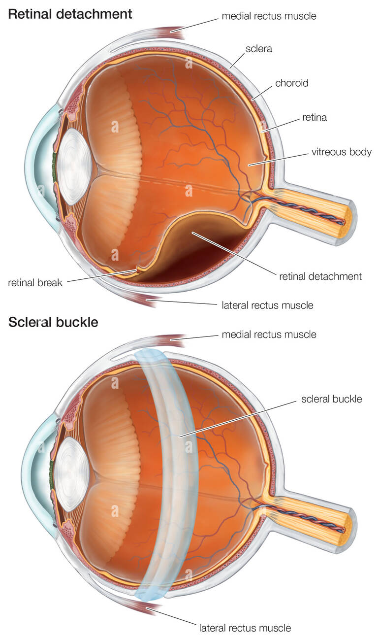
The retina is a layer located at the back of the eyeball containing light sensitive cells that send impulses to the brain to produce vision. Often, this layer of the eye can become damaged causing a retinal detachment. A retinal detachment occurs when the retina is pulled from its natural position at the back of the eye. One of the many ways to treat a retinal detachment is through the use of a surgical technique known as a scleral buckle.
What is a Scleral Buckle?
A scleral buckle is a surgical procedure that has been widely used for over six decades. This surgical technique has been favorable among surgeons throughout the years due to few complications and little to no need for post – operative positioning. The scleral buckle is a band typically made of silicone that is attached to the sclera, also known as the white part of the eye, at the site of the detachment. This “buckle” places an indentation on the eye wall allowing extra support to the retina by bringing the eye wall closer to the area of the detachment. Think of it like placing a rubber band around a balloon. The scleral buckle will remain around the eye permanently to aid in the prevention of future retinal detachments.

When is a Scleral Buckle Recommended?
The most common surgery for retinal detachment repair is a vitrectomy, where vitreous jelly is removed from inside the eye through small incisions in the sclera. However, there are advantages to repairing a retinal detachment with a scleral buckle, either alone or in combination with a vitrectomy.
There are many considerations your retina specialist must take into account when determining if a scleral buckle is the correct surgical option to treat your retinal detachment. Some of these considerations include:
- Age – Younger patients who have not undergone cataract surgery may benefit from a scleral buckle. Unlike other surgical procedures, scleral buckles do not accelerate the formation of a cataract.
- Location – The location of the tear/detachment plays a large factor in determining if a scleral buckle is the best option of surgical repair.
- Multiple breaks – Patients with multiple retinal tears or a large detachment in the eye would benefit from a scleral buckle. The scleral buckle would provide support to these areas as well as repairing your detachment. Additionally, patients with excessive areas of thin retina, known as lattice degeneration, would benefit from a scleral buckle due to it providing support to the entire retina.
- High Myopia – Patients who are significantly nearsighted would benefit from a scleral buckle versus other surgical interventions. Scleral buckles in these patient’s support the naturally tear-prone peripheral retina in myopic individuals.
- Scar Tissue on the Retina – Some cases of retinal detachment present with scar tissue on the retina. These ‘membranes’ of scar tissue pull the retina away from the eye wall and increase the risk for re-detachment after surgery. By utilizing a scleral buckle in these cases, the eye wall can be moved towards the retina, relieving the tractional forces caused by the scar tissue.
- Recurrent Retinal Detachment – Vitrectomy surgery fails to permanently reattach the retina in ~15% of cases. When a retinal detachment recurs, there may be hidden retinal breaks or scar tissue that prevented successful reattachment. In these cases, adding a scleral buckle helps to resolve untreated tears or scar tissue, allowing for permanent retinal attachment and preservation of vision.
How Is a Scleral Buckle Procedure Performed?
After you and your retina specialist have determined that a scleral buckle is the best surgical approach for treating your retinal detachment, it’s time to go to surgery. Due to continuing advancements in surgical techniques, this procedure can be performed safely in an outpatient surgical center. Scleral buckle placement can take anywhere between 45 minutes to one hour. Once you arrive to the surgery center, you will be given IV sedation and localized numbing. Dilating drops will be placed into the eye to widen the pupil allowing a full view to the back of the eye. A lid speculum is then placed into the eye, allowing it to remain opened during the procedure, and a drape is placed over the non-surgical eye. An incision is made in the outer layer of the eye (sclera) and the buckle is stitched around the circumference of the eye. To further prevent the detachment from recurring your surgeon may opt to do one of the following:
Cryopexy: A procedure in which extreme cold is applied to the outer surface of the eye to freeze it. This freezing process allows scar tissue to form and seal a break in the retina
Laser Photocoagulation: During this procedure a laser beam is applied to the area surrounding the tear or detachment causing a burn. This creates scar tissue to stop fluid from leaking into the retina.
Additionally, any residual fluid remaining behind the retina will be drained during the procedure and antibiotic drops will be placed into the eye to prevent infection after surgery. An eye patch will be placed over your eye, and you’ll ‘ll be instructed to follow up in the clinic the following day.
What Are the Expected Outcomes Following Surgery?
Once the procedure is complete, the patient will be allowed to return home the same day. You may feel some discomfort the day of your surgery and a few days following. However, prolonged discomfort is not expected. Redness, tenderness and some swelling of the eye is expected for a few weeks after surgery. Recovery for a scleral buckle procedure can take up to four weeks. During this time, you will also be placed on a drop regimen to prevent infection or inflammation in the eye. You may also be instructed to wear an eye patch during the day or at night to keep the eye protected during your recovery process. Additionally, it will be necessary to follow-up with your surgeon throughout your recovery process to ensure your eye is properly healing. It Is important to follow the guidelines given to you by your surgeon to ensure you do not harm the eye. A few dos and don’ts after having a scleral buckle include:

- Do not drive until you are cleared by your retina surgeon
- Do follow all post-operative care instructions including positioning requirements and use of medicated drops
- Do not fly if a gas bubble is placed in the eye.
- Do not lift heavy objects until you are cleared by your retina surgeon
Risks Factors
Proceeding with a scleral buckle typically yields positive results. However, as with all surgeries, there are risks that must be taken into account. Complications, although minimal, can occur with a scleral buckle procedure. These complications include:
- Infection
- New retinal tears
- Repeated detachments
- Double vision
- Acceleration of a cataract if a gas bubble is placed in the eye
- Elevated intraocular pressure
- Bleeding
Additionally, if you have undergone previous eye surgeries causing scar tissue, a scleral buckle may not initially treat your detachment. In the event this occurs, the procedure may have to be repeated so scar tissue can be removed.
It is important that you contact your retina surgeon if you are experiencing an increase in pain, swelling, decrease of vision, or bleeding after undergoing this procedure.
Dr. Anita Shane at Venice Retina is trained in diagnosing and treating retinal detachments using a scleral buckle technique. If you are experiencing any signs or symptoms related to a retinal detachment, call us for an appointment to discuss your surgical options!
