What is an Eye Floater?
You may have heard of a close friend or family member speaking about the floaters they experience in their eye, but what exactly does that mean? Floaters are exactly what their name represents – small dark spots that float in and out of your vision. Many people often describe their floaters as black specks, threads, or spider webs in their vision. Some even mistake their floaters for a small bug that is encroaching towards them!
Floaters can range in size and can cause significant interference in activities of daily living. When they happen suddenly, floaters can also be very alarming. It is important that if you are experiencing floaters, especially if they happen acutely, to have a dilated eye examination as there can sometimes be an underlying problem that requires attention. On the other hand, it is important to note that floaters are a very common symptom of adult patients and actually occur due to normal age-related change that is happening inside of the eye. To better understand exactly what floaters are and where they come from, let’s first understand the anatomy of our eyes.

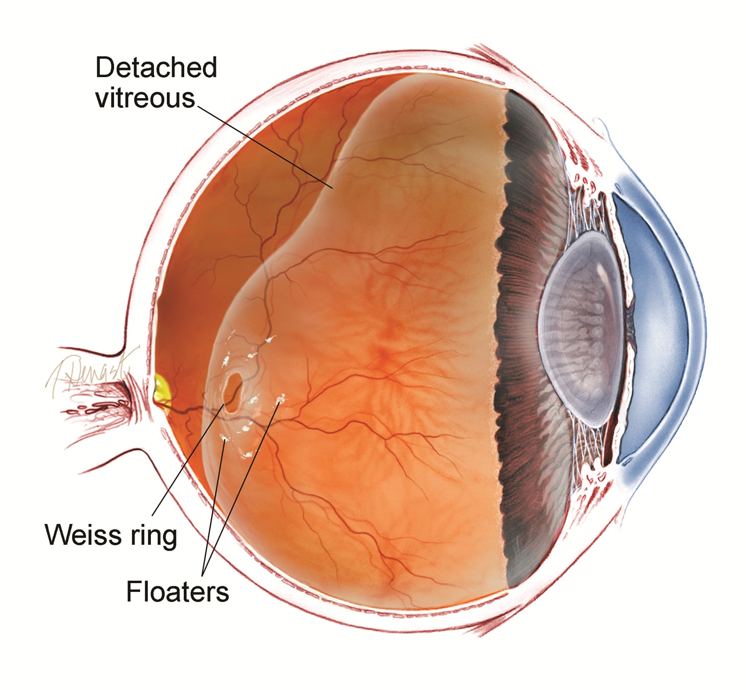
While small in size, our eyes are constantly performing extremely intricate tasks in order to promote vision. The inside of the eye is filled with a clear substance called the vitreous humor, and the very back of the eye is referred to as the retina. The vitreous humor is completely attached to the retina through small, microscopic fibers made of collagen. The retina itself is a thin layer of light-sensitive tissue that lines the inner surface of the back of the eyeball and transmits visual signals to our brain through the optic nerve. Without a proper functioning retina, our brain would not be able to interpret any visual impulses, and thus, we would not be able to see. For visualization purposes, if you envision the eye as a small, inflated basketball, the air inside of the basketball would represent the vitreous humor, and the interior rubber lining of the basketball itself would represent the retina.
When we are born, the vitreous humor is more gel-like in appearance. As we age, however, it continuously liquefies into a more fluid-like solution, resembling the appearance of water. Over time, the vitreous humor naturally begins to separate from the retina, breaking apart the small microscopic connections that once attached the two together. This is referred to as a PVD, or posterior vitreous detachment. Once used as an adhesive to attach the vitreous humor to the retina, the collagen fibers are now floating around inside of the eye itself, creating the floaters that you see in your vision!
It is important to note that a PVD is common – it spontaneously happens to everyone as we age. You can think of it as a natural progression in the life cycle of the eye. Most often directly related to age, a PVD can also occur (though less commonly) as a result of trauma to the eye. While floaters are more frequently related to a PVD, they can also arise from other, more serious problems in the eye which is why it is important for any floaters – especially new ones! – to be examined by your retina specialist. As the vitreous jelly is separating from the retina, it can sometimes peel off with enough force and traction to pull on the very delicate retina, creating a retinal tear or detachment. These conditions require immediate medical attention and can cause severe vision loss if not treated in time.

What Happens During My Eye Exam?
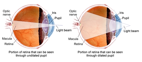
So, you’ve made an appointment quickly for your new floaters – fantastic! We recognize how scary changes in your vision can be so we want you to feel at ease in knowing what will occur beforehand. Once you come into the office, your visual acuity will be checked on an eye chart, your peripheral vision will be tested, your intraocular pressure (IOP) will be taken, and your eyes will be dilated. You may also have photos taken of your retina called OCT’s (Optical Coherence Tomography) or even a video taken of your floaters ensuring that we objectively document exactly what you see! Next, your retina specialist will take an in-depth look inside your eye to evaluate every square millimeter of the retina, ensuring that there are no tears or detachments responsible for the bothersome floaters you are experiencing.
Will My Floaters Harm My Eye?
Our office recognizes how scary and troublesome new floaters can be – our eyes are delicate and any changes are cause for immediate concern! Your floaters themselves are not dangerous to the eye. Made up of organic collagen fibers, your floaters themselves are not harmful, toxic, or damaging to the eye. Instead, they are a cause for frustration as they wisp around the vitreous humor and impede your vision. What is dangerous, however, are the problems that are hidden behind new floaters, such as an underlying retinal tear or detachment. Your floaters should be evaluated quickly with a qualified ophthalmologist or eye doctor to ensure there are no hidden, vision-threatening problems happening.
Are There Any Ways To Get Rid Of My Floaters?
Yes – there are absolutely ways to help! If your floaters have been present for at least 6 months and are causing significant interference with your vision or activities of daily living, you may be a good candidate for surgery to remove your floaters completely.
The surgery is called a Pars Plana Vitrectomy (PPV) and takes place in an outpatient surgery center. During the procedure, your eye is numbed completely and the vitreous humor inside the eye is removed. Remember that the vitreous humor contains all of the collagen fibers that clumped together and are causing your floaters, so this surgery is guaranteed to remove your nagging floaters altogether. At the time of surgery, the vitreous humor is replaced with an artificial saline solution and then over the course of 24-48 hours, it is again replaced with the normal, clear fluid constantly produced inside the eye.

This procedure only takes about 15 minutes and occurs under local anesthesia. This is a very common retina procedure without any significant postoperative restrictions, and your floaters are immediately resolved.
Always remember that there is another option – observation. While effective in removing floaters, both surgery and the use of anesthesia each have inherent risks associated with them so patients may feel more comfortable with continued monitoring to see if there are improvements in their floater symptoms without requiring surgery. Dr. Shane will be sure to provide a thorough and detailed discussion regarding the risks, benefits, and alternatives to treatment as it relates to you and your specific needs!
Will My Insurance Cover A Floater Removal Procedure?
Dr. Shane is in network with most major insurance providers and they typically cover your floater treatment. Of course, this is subject to your insurance plan and its associated copay and deductibles. When you contact us to schedule an appointment, feel free to inquire with one of our qualified staff members for more information!
Common Retina Care Questions
How Long Is The Recovery?
One of the benefits of a PPV is that there are no significant restrictions following the surgery itself, and the recovery period is rather short. Our office would follow you postoperatively to ensure that the eye is healing as it should without any problems, but you would be able to return to work, school, or other obligations immediately (if you feel comfortable).
I Have Another, Underlying Eye Problem (I.E. Glaucoma, Macular Degeneration, Etc.) – Do I Still Qualify For Floater Removal?
Even if you have an underlying eye diagnosis that you receive treatment for, such as Glaucoma or Macular Degeneration, you can still qualify for surgery to treat your nagging floaters. Dr. Shane will be sure to address any questions or concerns, and recommend a plan for you!
How Many Vitrectomy Surgeries Has Dr. Shane Performed?
Hundreds – if not thousands! Interestingly enough, a vitrectomy is commonly the very first step in any retina surgery. For example, before repairing a detached retina, you would often start the surgery with removing the vitreous humor! Dr. Shane is an accomplished surgeon and has years of experience and expertise in all retinal surgeries, specifically vitrectomies.
What Should I Do Next?
If you have been experiencing floaters, especially new ones, please contact our office for an evaluation! Once again, sometimes floaters can be a sign of an underlying retinal tear or detachment, which require immediate medical care. If not treated in time, they can cause irreversible vision loss. We want the best for you and your eye health, so don’t delay!
