What is a Macular Pucker?
Also known as an “Epiretinal Membrane”
An epiretinal membrane is an extremely common diagnosis for patients ages 50 and older – but what exactly is it? You may have heard it by a couple of commonly used names, such as a retinal wrinkle, macular pucker, or cellophane maculopathy, though they are all used interchangeably and mean the same thing! To better understand exactly what this means, however, we first need a quick lesson in the anatomy of our eyes. The retina is the delicate, light-sensitive tissue that lines the back of our eyes. The retina collects light that enters our eye through the lens and transmits these signals to the brain through the optic nerve. In the middle of our eyes is the vitreous humor – a jelly-like substance that fills the space of the eye between the lens and the retina. You can think of the vitreous humor in the same way that you think of air filling the middle of a soccer ball.

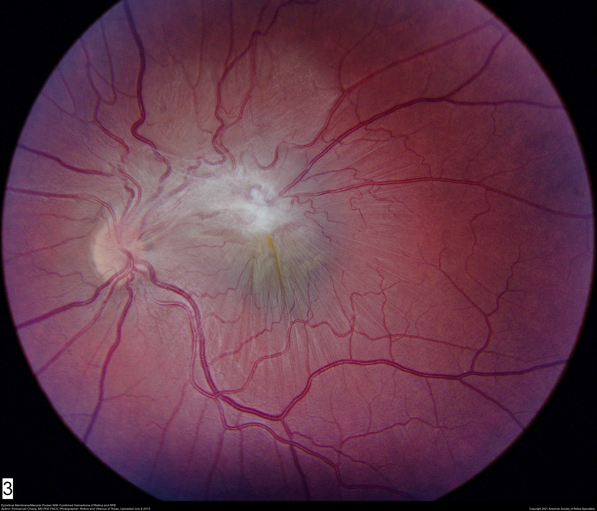
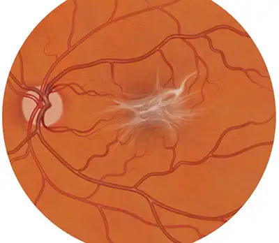
Now, the very center-most portion of the retina is referred to as the macula. This is the location where an epiretinal membrane, or macular pucker, forms. The macula measures only about 5 millimeters in length, but is responsible for your intricate, central vision. A macular pucker occurs when a microscopically thin sheet of tissue forms on top of the macula, pulling on the retina and wrinkling it, thus, causing a disturbance in vision.
What Are The Symptoms Of A Macular Pucker?
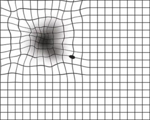
 Sometimes, patients who have a macular pucker may be asymptomatic. However, if it progresses to the point that your epiretinal membrane tugs on the retina, it will begin to move the delicate photo receptor cells that aid in vision, causing a variety of symptoms. If you have an epiretinal membrane or a “wrinkle” in the retina, your symptoms will appear gradually (typically months or years) and painlessly. You will likely experience slight distortion in your vision – you may see straight lines as wavy or words in a sentence as blurred. You may struggle reading fine print, such as in your favorite book or closed captioning on your television show. Most people with a macular pucker will not experience severe vision loss.
How Did I Get A Macular Pucker? Is It Something I Did? Is It Genetic?
While sometimes patients may not even know they have a macular pucker until receiving a dilated eye exam. While quite common, it is important to note that epiretinal membranes are nonetheless not a part of the normal eye anatomy. As mentioned earlier, the middle of our eye is filled with vitreous humor – a jelly-like substance that is attached to the retina at the back of the eye through microscopic tethers. As we age, this jelly spontaneously and naturally separates from the retina, a development called posterior vitreous detachment (PVD). This process often leaves behind some of the small adhesive cells on the macula. These cells can bunch together, acting like scar tissue, spreading themselves across the retina and eventually contracting, forming your retinal wrinkle and causing visual distortions.
There are no studies to suggest that there is a genetic predisposition to having an epiretinal membrane, but there are some risk factors that may make you more likely to develop one. Some risks factors include, but are not limited to:
- History of Eye Surgery or Retinal Laser Treatment(s)
- History of a retinal tear or retinal detachment
- Ocular trauma
- Other retinal comorbidities, such as diabetic retinopathy
- Intraocular bleeding or inflammation
If you have any of the above conditions, then you may be more likely to develop a macular pucker.

How Is An Epiretinal Membrane Diagnosed?
If you suspect you may have an epiretinal membrane, or are experiencing any of the associated symptoms, you should seek a dilated eye exam with an ophthalmologist, specifically a retina specialist. At your appointment, your medical history will be obtained, visual acuity will be checked, intraocular pressure measured, and pupils dilated. Additionally, specialized images of the back of your retina will be taken, called OCT’s (Optical Coherence Tomography). This is a noninvasive method to take high-resolution photos of your retina, allowing your retina specialist to view your epiretinal membrane in its totality. Next, your retina specialist will examine your eyes, review your images, and discuss with you the findings of your exam, as well as any risks, benefits, and alternatives to treatment.
How Is A Macular Pucker Treated?
As mentioned previously, some patients may not even recognize that they have an epiretinal membrane as most cases are mild and cause only limited blurring or distortion in vision. Epiretinal membranes progress very slowly, and are not an immediate threat to irreversible vision loss – it can sometimes take years for a patient to recognize distortion in their vision! Thus, many times it is safe to simply observe these patients without pursuing further treatment.
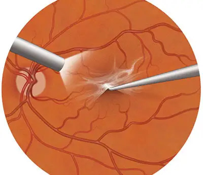
However, in those cases where the epiretinal membrane continues to cause significant distortion in vision, interfering with the activities you enjoy on a daily basis, your retina specialist may recommend surgery to remove the epiretinal membrane. This is a commonly performed surgery that takes place at an outpatient, same-day surgery center under local anesthesia. During surgery, your retina specialist will remove the vitreous jelly inside of your eye, a procedure referred to as vitrectomy. If you have any floaters in that eye, they will be eliminated during this phase of the vitrectomy. Once the jelly is removed, microscopic tweezers are used t0 peel the membrane off of the surface of the macula, removing the epiretinal membrane in its entirety. This whole procedure takes about 30 minutes.


The recovery following surgery has only minimal restrictions. You will use eye drops prescribed to you following surgery and be evaluated postoperatively to ensure that you’re healing properly. Your vision is expected to continuously improve as your eye adjusts back to its baseline. Dr. Shane will be sure to discuss all of the risks, benefits, and alternatives with you, as well as answer any questions you may have during your consultation.
Is This A Common Procedure?
Yes! Dr. Shane has performed thousands of vitrectomy procedures and epiretinal membrane removals. This is one of the most frequently performed retina procedures at Venice Retina!
Is Surgery Covered By My Health Insurance?
Dr. Shane is in network with most major insurance providers and, thus, this procedure is covered by your health insurance plan. Our office will obtain an authorization with your insurance prior to surgery and the price of the procedure would be subject to any preexisting deductibles or copay unique to your specific plan. If you have any questions about this, feel free to contact our very qualified billing staff for any further assistance!
What Should I Do Next?
If you feel as if you have some of the symptoms discussed previously or have a predisposing factor to potentially develop a macular pucker, please schedule a consultation with Dr. Shane!
