Optical Coherence Tomography (OCT)
A Non-Invasive Diagnostic Imaging Device
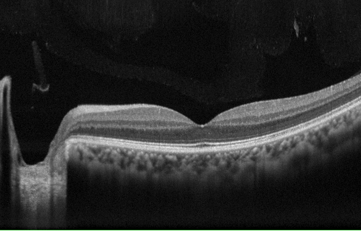
Optical coherence tomography (OCT) is a non-invasive diagnostic imaging device that utilizes light waves to capture an image of your retina. An OCT allows the ophthalmologist to see the distinct cross-sectional layers of the retina. These images provide guidance for the ophthalmologist so they can better manage retinal treatment plans. This form of imaging can capture a number of retinal conditions including Age-related macular degeneration and diabetic changes. The OCT is also able to diagnose conditions involving the optic nerve.
How Does Optical Coherence Tomography Work?
OCT imaging is similar to an ultrasound or an X-ray. However, an OCT uses light waves to produce an image, rather than sound waves or radiation. The OCT can take an image of the retina, optic nerve or other parts of the eye such as the cornea. The camera will send slices of light through the eye and turn the slices sideways to produce an image. OCT allows the ophthalmologist to see the individual layers of the retina, thickness of the optic nerve and/or thickness of the cornea.
When Is an OCT Recommended?
Determining when a patient needs an OCT is a critical decision. Most new patients undergo optical coherence tomography imaging. This gives your ophthalmologist a baseline retinal status to compare during future imaging.
Patients who are symptomatic experiencing changes in vision or loss of vision should undergo periodic OCT imaging. This will allow your ophthalmologist to monitor the progression of your condition. On the other hand, patients who have excellent visual acuities and are asymptomatic may not benefit from routine OCT imaging.

How Is an OCT Performed?
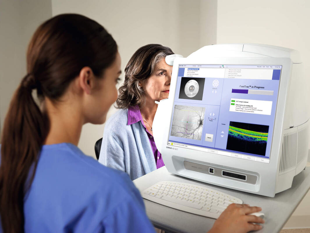
OCTs are taken at the beginning of your exam so the images can be reviewed by the time you see the eye doctor. Dilating drops may or may not be required to widen your pupil prior to imaging. Once you are seated at the machine, you will be positioned on the chin rest to help keep your eye steady. There is a light in the back of the machine that you will be instructed to look at while your eye is being scanned. While the image capture takes less than 10 seconds, the entire process of positioning and scanning will take anywhere between five to ten minutes.
Ophthalmologists utilize OCT imaging to diagnose a wide variety of conditions in the retina, optic nerve, and anterior segment. These conditions include:
Retina

- Macular hole
- Macular Pucker
- Macular Edema
- Age-related macular degeneration
- Central Serous Retinopathy
- Diabetic Retinopathy
Optic Nerve
The OCT is also capable of evaluating optic nerve disorders by viewing the nerve fiber layer to evaluate conditions such as:
- Glaucoma
- Optic neuritis
- Alzheimer’s disease
- Non-glaucomatous optic neuropathies
Anterior Segment

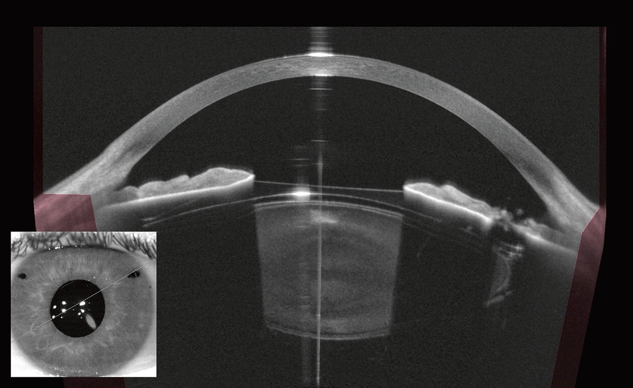
The anterior segment OCT utilizes higher wavelengths on the OCT machine. The increase in wavelengths allows for greater absorption and less penetration so the front of the eye can be captured and measured. This allows for images of the cornea, anterior chamber and iris to be visualized.
What Are Limitations to OCTs?
Due to the OCT utilizing light waves, limitations to its reliability must be taken into consideration when ordering this diagnostic tool. If a patient presents with blood in their vitreous or if they have a dense cataract, the image will not be clear. These types of opacities will interfere with the OCT imagining, limiting its quality.
Additionally, patient cooperation is a necessity to utilize this diagnostic tool. If a patient moves during the testing the quality of the image can be altered. However, with continued advancements in OCT technology, images are increasingly possible even in patients who cannot remain absolutely still.
Contact Us!
Venice Retina prides itself on using advanced imaging techniques to manage retinal conditions. If you are experiencing any changes in your vision, contact us today to receive the appropriate diagnostic imaging!
